How to Count Cells Using Stereology: A Step-by-Step Guide
Counting cells reliably is not about tallying them one by one under the microscope: it requires systematic random sampling and the optical disector, two principles that guarantee an unbiased result regardless of the size, shape, or orientation of the cells being observed.
Why You Can't Just "Count Cells" Under a Microscope
The Problem of Bias in Manual Counting
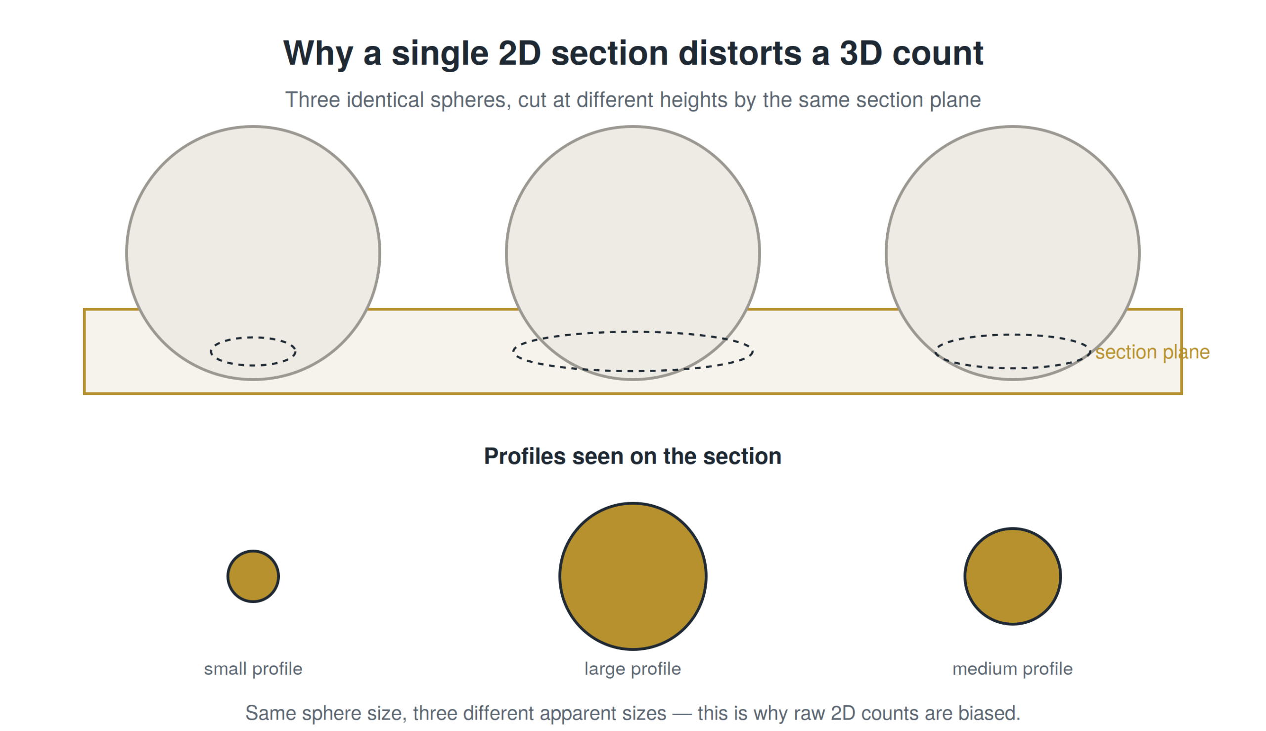
A visual count, even performed carefully by an expert, almost always produces a skewed estimate. This isn't a matter of the observer's rigor — it's a geometric bias inherent to how a two-dimensional tissue section represents a three-dimensional structure. The same cell can appear on several consecutive sections, or conversely never be visible at all if it falls between two cutting planes. Without a corrective method, the resulting count depends as much on section thickness and cell size as on the actual number of cells present.
Why Cell Size and Orientation Distort Naive Counts
This problem has been known for nearly a century: as early as 1925, statistician Sven Wicksell demonstrated that a three-dimensional structure observed through a flat section systematically overestimates larger objects, since they are statistically more likely to be "hit" by the cutting plane than smaller ones. This bias, now known as the "Wicksell effect" or size-frequency bias, explains why two researchers counting the same section with different methods can arrive at significantly different results.
The Core Principle: Unbiased, Systematic Random Sampling
What "Unbiased" Actually Means in Stereology
In stereology, a method is considered "unbiased" when it relies on no assumption about the size, shape, or orientation of the objects being counted. Unlike density-based counting methods, which assume a regular distribution of cells, unbiased methods work regardless of how irregular the actual tissue is. This absence of assumptions is precisely what makes results comparable across studies and laboratories.
Systematic Uniform Random Sampling (SURS) Explained Simply
SURS involves choosing a random starting point within the tissue, then sampling subsequent sections at fixed, predefined intervals. This combination of randomness (the starting point) and system (the fixed interval) ensures that every part of the tissue has an equal chance of being included in the sample, without the researcher having to make subjective choices about which areas to observe.
Step-by-Step: How to Count Cells with the Optical Fractionator
The optical fractionator, developed by Peter Sterio in 1984 and later refined by Hans Jørgen Gundersen and colleagues, is today the gold-standard method for estimating the total number of cells in a tissue structure.
Step 1 — Define Your Region of Interest (ROI)
Before any counting begins, the anatomical structure under study (a brain nucleus, a cortical layer, an entire organ) must be precisely outlined on each section. This delineation should follow reproducible anatomical criteria, ideally defined in advance and applied identically by every observer in a given study.
Step 2 — Set Up Systematic Random Sampling of Sections
Across the full series of sections covering the structure, a known, regular fraction is selected (for example, every fifth or every tenth section), starting from a random point among the first sections. This fraction becomes the first factor in the final formula.
Step 3 — Place Counting Frames and Disectors
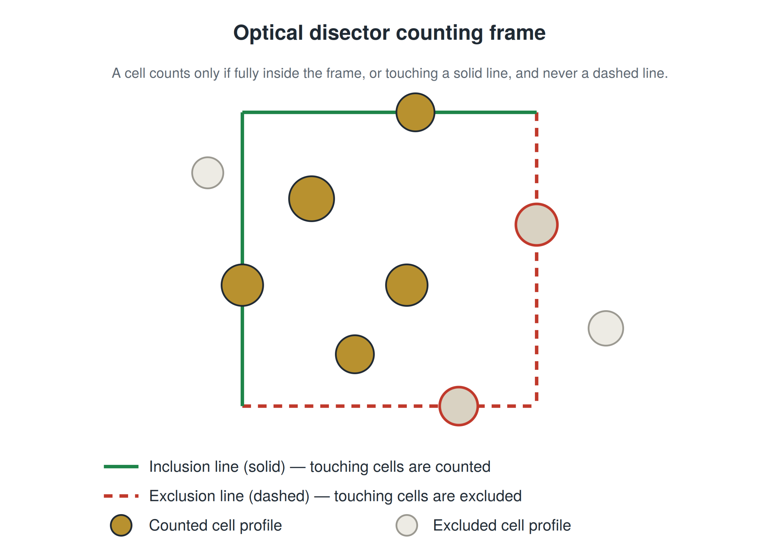
On each sampled section, counting frames are positioned according to a systematic random grid. Each frame defines an "optical disector": a small three-dimensional volume within the section's thickness, inside which cells are actually counted, with a guard zone at the top and bottom of the section to avoid sectioning artifacts.
Step 4 — Apply the Counting Rules
A cell is only counted if: it is entirely visible within the disector volume, it does not touch the frame's exclusion lines, and its uppermost point comes into focus for the first time within the sampled volume. These rules, formalized by Gundersen as early as 1977, mathematically eliminate the size-frequency bias described by Wicksell.
Step 5 — Calculate the Total Cell Number
The total number of cells (N) is obtained by multiplying the number of cells counted within the disectors by the inverse of each sampling fraction applied.
N = ΣQ⁻ × (1/ssf) × (1/asf) × (1/tsf)
where ΣQ⁻ is the total number of cells counted, ssf the section sampling fraction, asf the area sampling fraction covered by the counting frames, and tsf the thickness sampling fraction of the disector relative to the full section thickness.
Simplified example: if you count 200 cells (ΣQ⁻), with every tenth section sampled (ssf = 1/10), counting frames covering 1/50 of the total surface (asf = 1/50), and a disector using 1/2 of the section thickness (tsf = 1/2), then N = 200 × 10 × 50 × 2 = 200,000 estimated cells.
Tools You'll Need
Microscope Requirements
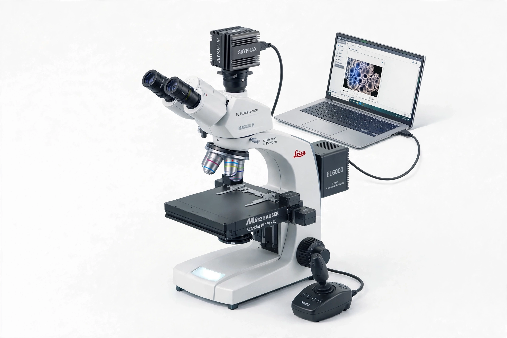
Manual stereological counting requires a microscope equipped with a motorized X-Y stage, a Z-axis controller with micrometer precision, and ideally a camera connected to a computer for software-driven counting.
Software Options
StereoInvestigator (MBF Bioscience) dominates the academic market, but alternatives exist, including some open-source options, particularly for labs that already own a motorized microscope but want to reduce licensing costs.
Common Mistakes That Bias Your Cell Counts
- Undersampling: too few sections or frames counted, which increases the coefficient of error without the user necessarily noticing
- Poorly defined region of interest: vague or inconsistent anatomical criteria from one section to the next distort the very structure being counted
- Ignoring inclusion/exclusion rules: counting a cell that touches an exclusion line, or missing one that touches an inclusion line
- Confusing density with total number: reporting cell density without accounting for volume differences between study groups, which can mask or fabricate a biological effect
Optical Fractionator vs. Other Counting Methods
| Method | Principle | Biais |
| Optical fractionator | Direct counting within a sampled 3D volume | Unbiased |
| Isotropic fractionator | Dissociation of tissue into a homogeneous suspension, counting using aliquots | Unbiased, but loses spatial information |
| Density-based counting (2D profile) | Counting cell profiles on thin sections, without a count chamber | Biased (Wicksell effect) |
FAQ
Is stereology cell counting time-consuming?
Yes, a rigorous count using the optical fractionator can take several hours per sample depending on the size of the structure and the desired level of precision. That's the trade-off for obtaining an unbiased estimate.
What sample size do I need for reliable estimates?
Sample size depends on the desired coefficient of error. In practice, researchers typically aim to count between 100 and 200 cells per structure, spread across 8 to 12 sections, to achieve an acceptable sampling error.
Can cell counting be automated with AI/deep learning?
Deep learning approaches, particularly those based on image segmentation architectures like U-Net, are increasingly used to assist or speed up certain steps of stereological counting. These tools are generally used alongside classical stereological rules rather than as a full replacement.
Do I need special software to count cells with stereology?
Dedicated software isn't strictly required, but it greatly simplifies compliance with counting rules, disector coordinate management, and automatic calculation of total cell numbers.
Learn More: Structured Training in Unbiased Stereology
This guide covers the essential principles of stereological cell counting, but reliable practice requires guided training with hands-on application on real samples.