Optical Fractionator Method in Stereology
The optical fractionator is the gold-standard stereological method for estimating the total number of biological objects — cells, neurons, specific cellular structures — within a tissue, combining multi-level systematic random sampling with disector-based counting, without any assumption about the size or shape of the objects being counted.
Origin and Principle of the Method
From the Disector to the Optical Fractionator
The optical fractionator was developed by West, Slomianka, and Gundersen in 1991, combining two earlier advances: the disector principle, introduced by Sterio in 1984, and the systematic fractionator principle, which allows a total number to be estimated from a known, representative fraction of the tissue. This combination made the method both mathematically rigorous and practical for routine laboratory use.
Why "Fractionator"?
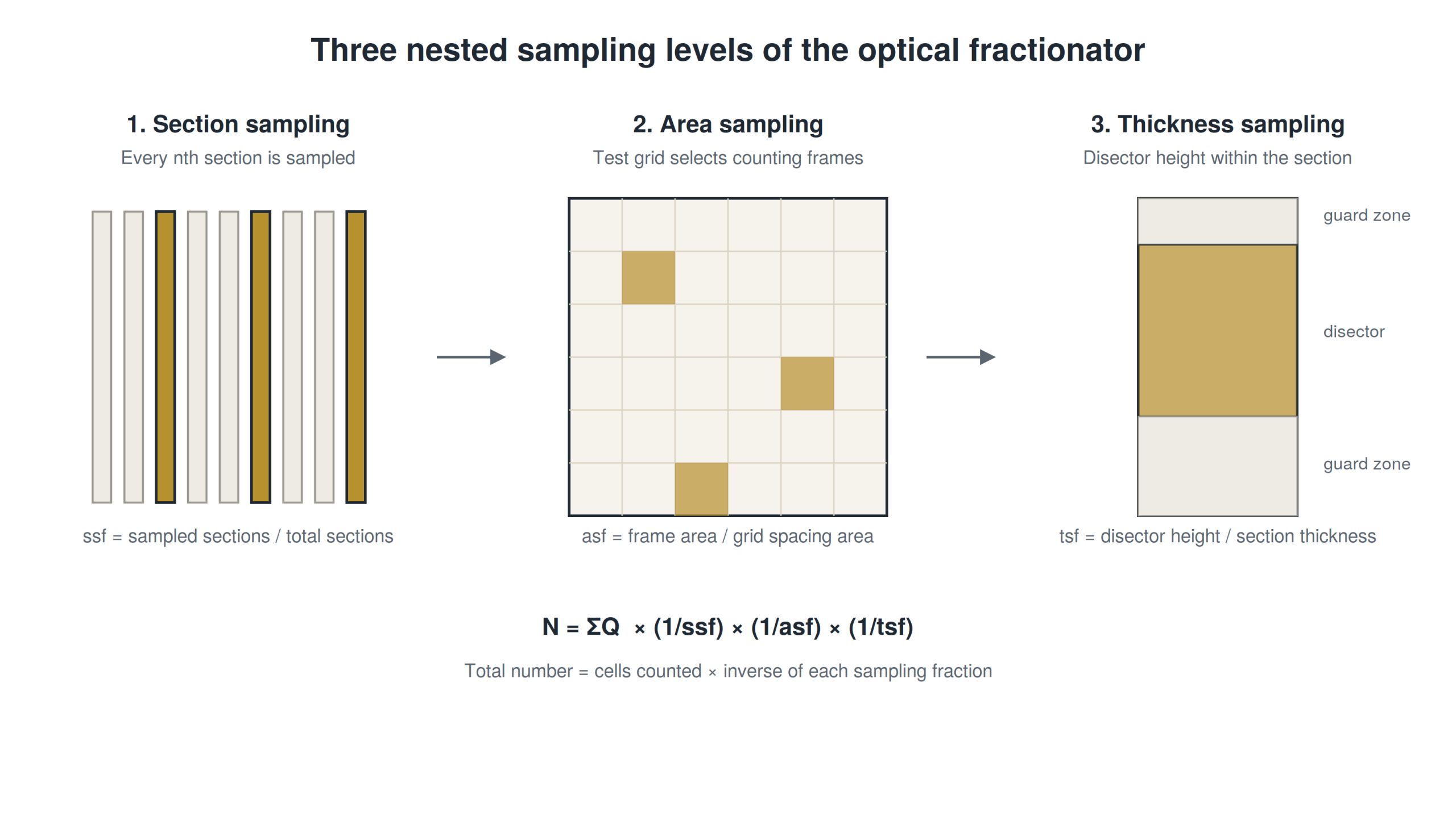
The name comes from the method's central principle: rather than counting an entire structure, objects are counted within a known fraction of its total volume, at three nested levels — section fraction, area fraction, thickness fraction — and the total number is obtained by multiplying by the inverse of each of these fractions.
The Detailed Protocol, Step by Step
Step 1 — Delineate the Region of Interest on Each Section
The structure under study must be outlined on each sampled section using fixed, reproducible anatomical landmarks, applied identically throughout the study. Any variation in this outline introduces a source of error unrelated to the stereological counting itself.
Step 2 — Define the Section Sampling Fraction (ssf)
Across the full series of sections covering the structure, every nth section is systematically selected, starting from a section chosen randomly among the first n sections. This fraction forms the first of the three factors in the final formula.
Step 3 — Define the Area Sampling Fraction (asf)
On each selected section, a systematic random test grid determines the positions where counting frames will be placed. The area sampling fraction corresponds to the ratio between the area of a counting frame and the area covered by the test grid.
Step 4 — Define the Thickness Sampling Fraction (tsf)
Within each counting frame, the optical disector uses only part of the section's total thickness, excluding a guard zone at the top and bottom to avoid sectioning artifacts. The thickness sampling fraction is the ratio between the disector's height and the total section thickness after processing.
Step 5 — Apply the Disector Counting Rules
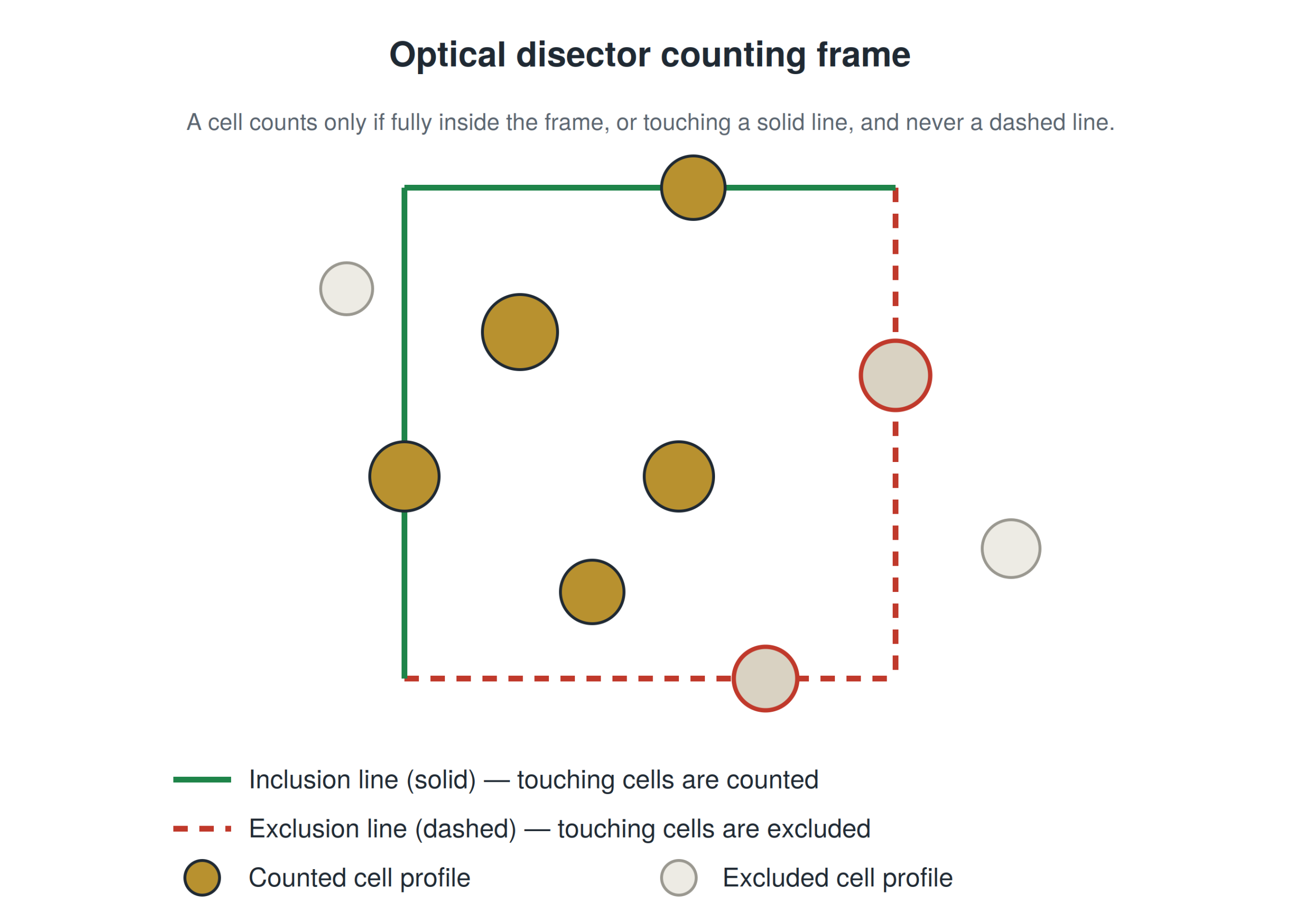
An object is only counted if it is entirely contained within the disector volume, does not touch any of the counting frame's exclusion lines, and its uppermost point comes into focus for the first time within the sampled zone. These rules, formalized by Gundersen as early as 1977, guarantee freedom from bias related to object size or shape.
Step 6 — Calculate the Estimated Total Number
The total number of objects (N) is obtained using the following formula:
N = ΣQ⁻ × (1/ssf) × (1/asf) × (1/tsf)
Detailed example: a study examines a structure covered by 40 serial sections. Every fifth section is sampled (ssf = 1/5, i.e. 8 sections analyzed). On each section, the counting frames cover 1/40 of the structure's total area (asf = 1/40). The disector uses 10 µm out of a total post-processing section thickness of 25 µm (tsf = 10/25 = 2/5). If 150 objects are counted in total (ΣQ⁻ = 150), then: N = 150 × 5 × 40 × 2.5 = 150,000 estimated objects.
Estimating Precision: The Coefficient of Error
An optical fractionator estimate should always be reported alongside its coefficient of error (CE), which quantifies the share of variability due to the sampling design itself, independent of true biological variability between the subjects studied. A CE that is too high signals insufficient sampling and calls for revisiting the chosen fractions before treating the results as reliable.
Common Pitfalls and How to Avoid Them
- Poorly positioning the disector's guard zones: a guard zone that's too thin doesn't adequately protect against compression artifacts
- Confusing inclusion and exclusion lines: a common beginner mistake that silently reverses the direction of the bias
- Tissue shrinkage: the tsf must be calculated using section thickness after processing, not the thickness set at the microtome
- Over-interpreting a ΣQ⁻ that's too low: fewer than 100 to 150 total objects counted generally leaves the coefficient of error too high for a solid statistical conclusion
Optical Fractionator and Stereology Software
In practice, most laboratories use dedicated software (StereoInvestigator being the most widely used) to drive the microscope's motorized stage, automatically generate the test grid, and directly calculate N and its coefficient of error from the entered counts — greatly reducing the risk of manual calculation errors.
For a broader overview of the methodological context, see our "Unbiased Stereology Tutorial". For the step-by-step practical application to cell counting, see "How to Count Cells Using Stereology".
FAQ
Does the optical fractionator work for any type of tissue?
Yes, in principle: the method makes no assumption about the shape or distribution of the objects being counted. In practice, tissue quality affects how easy the method is to apply, but not its theoretical validity.
What's the difference between the optical fractionator and the physical disector?
The original physical disector requires comparing two physically separated adjacent sections. The optical fractionator uses a single tissue block and moves optically through its thickness, making it much faster and less destructive to the sample.
How long does it take to apply this method to a sample?
This varies significantly depending on the size of the structure and the density of objects to count, but a rigorous count generally takes several hours per sample.
Can the optical fractionator be partially automated?
Some software now includes AI-assisted detection to propose a pre-count, which the user then validates or corrects manually.
Learn More: Structured Training in Unbiased Stereology
The optical fractionator formula is simple on paper, but its rigorous implementation requires guided practice.